LEVEL 3 (Form 4-5 & Foundation in Science)
- Aug 9, 2016
- 11 min read
LESSON 1: Prokaryote VS Eukaryote Cell


Source of image: http://www.e-nox.net/images/illustrations/cholerabacterium_full.jpg
CELL STRUCTURE AND FUNCTION
WHAT IS PROKARYOTIC CELLS ?
Basically, "pro" means before, and "karyote"means kernel, nucleus. They are so named because this type of cell lack a membrane-enclosed nucleus. Prokaryotes are single-celled organisms that are the earliest and most primitive forms of life on earth. They are the most abundant and diverse life-forms on Earth, plus, they present in a great numbers in the air, water and soil, as well as living in and on other organisms
THE STRUCTURE OF PROKARYOTE
Generally, prokaryotes are quite small. The average size of prokaryotes is 1.1-1.5 um wide and 2.0-6.0 um long. Try to imagine how tiny it is. That is why we cannot see it using our naked eyes. So as its shape, it is also has been determined. The most common 3 basic shapes are :

Source of image: Izyan Fartini, UPM
A rod-shaped bacterium is called bacillus while a spherical-shaped bacterium is known as coccus. Both of these can occur in pairs or chains. As for coccus they can actually form as clusters. Some bacteria are twisted into spiral, we called it spirilla if they are rigid but if they are flexible, we called it spirochete.
There are 4 major component of cells. Which is, plasma membrane, cytoplasm, DNA and ribosomes. Now we are focusing on prokaryotic cells first. Do not think of eukaryotic cells yet. As we are already mentioned, prokaryotic cell is a simple cell. Their inner part is not divided with any membrane. That is why prokaryote are known as a lack membrane-bounded organelles. Organelles means “little organ”. We will discuss more about organelles in eukaryotic cells after this.
Prokaryotic cells contain:

This is the picture of prokaryote cell.

Source of image: modified from "Prokaryotic cells: Figure 1" by OpenStax College, Biology, CC BY 3.0[1][1]https://www.khanacademy.org/science/biology/structure-of-a-cell/prokaryotic-and-eukaryotic-cells/a/prokaryotic-cells
THE STRUCTURES AND ITS FUNCTION
Prokaryotic cells are not as complex as eukaryotic cells. Here are the structures of the cell.


Source of image: http://www.asknature.org/images/uploads/strategy/47050f93e632116925dd813427b2767d/cell.jpg
Well, we know that prokaryotic cells do not have internal membrane. That is the main different between prokaryote and eukaryote. Do you still remember we discussed about the function of the structure? Eukaryote have a plasma membrane that separates the content of the cell from its surrounding. The plasma membrane is a phospholipid bilayer which is embedded with protein. So we can say that eukaryotic cells are compartmentalized. The compartment is typically called as organelle. Each of the organelles carry out their own specific function.
Eukaryotic cells are usually referring to plant and animal cells. Unlike prokaryotic cells, eukaryotic cells have numerous membrane bound organelles such as endoplasmic reticulum, Golgi apparatus, chloroplasts, mitochondria, and others. Let’s take a look.
THE STRUCTURES AND FUNCTION OF EUKARYOTIC CELLS


LESSON 2: Microbial Growth
This is important for you to know because you can use this info to prevent infection in everyday life. Ever heard sterilization? It is a process to kill or deactivate all forms of life either using chemical or heat.
Why it is important? To prevent the spread of infection because microbes can attach at any surface.
What sterilization has anything to do with microbial growth? The relevance is, a mesophilic microbe requires a suitable temperature for example 20 - 45 Celsius to grow. When we supply temperature more than 45 Celsius, we are able to kill the microbes instantly. In order to kill microbes, we must know its requirement so that we know their weaknesses. We also need to know their requirement if we want to culture good microbes & to maintain their growth so that they keep on producing end products that benefit us.
There are two types of microbial requirements:
Physical requirement
Chemical requirement
Let's check out what is the physical requirement for microbes shall we?
Physical Requirement
Temperature
pH
Osmotic pressure
Temperature
Microorganims are classified into three primary groups based on their preferred temperature range:
Psychrophiles : cold loving microbes (-10'C – 20'C, 15'C optimum temperature )
Mesophiles : moderate-temperature loving microbes ( 20'C – 45'C, 37'C optimum temperature )
Thermophiles : heat loving microbes ( 45'C – 60'C, 50'C-60'C optimum temperature)
*optimum temperature is the temperature which microorganisms grow at best and the most rapid rate.
pH
Microorganisms can be divided into three classes based on pH requirements.
Acidophiles : Microorganisms that able to tolerate with acidic environment (below pH 4). The optimum pH of Molds and yeast is generally below that of bacteria, usually about pH 5 to 6. Example of bacteria, Lactobacillus sp. These bacteria produce lactic acid, thus lower the pH. They are able to tolerate with mild acidity.
Neutrophiles : Microorganisms that grow at pH 5.4 to 8.5 include most human pathogens. Most bacteria grow best between pH 6.5 and 7.5 in neutral environment.
Alkaliphiles : Microorganism that loves alkali and grow at high pH between pH 7 to 12 or higher. Example bacteria, Agrobacterium which grows at pH of 12.
Osmotic pressure
Microorganisms obtain their nutrients in the form of solution from their surrounding that consist of water. Thus, water is the most important component for them as it is the main source of their food. They require 80-90% of water for growth.
Osmotic pressures have the effect of removing or transferring water in and out of the cell. There are three environments to describe the effect of osmotic pressure to the cell:

Hypertonic environment to the cell : When a microbial cell is living in a solution whose concentration of solutes is higher than in cell , the water in the cell will passes out to the environment with high solute concentration.
Plasmolysis : This osmotic loss of water causes (cell cytoplasm’s shrinks).
Isotonic: Net movement of microbes in & out of the cell are equal.
Hypotonic environment to the cell: If the environment has low in solute , water tends to enter the cell via osmosis. However, some microbes that have a weak cell wall may be lysed by such treatment, this condition is called osmotic lysis.
Chemical requirements
Macronutrients - require in large amount
Carbon - Carbon is the most important component besides water becuase carbon is needed for the structural backbone of living matter.Example of microorganisms that use carbon are called chemoheterotrophs (microorganisms that seeks for organic matter). However, microbes that are called as chemoautotrophs does not require organic matter for their energy source but they derive their carbon from carbon dioxide.
These elements below are necessary for microbes because these are important components to synthesize cellular material.
Nitrogen
Sulphur
Phosphorus
Trace elements - require in minute amount
Copper
Magnesium
Molybdenum
Zinc
Iron
These elements are essential for the functions of certain enzymes usually they are used as cofactors. Cofactors are non-protein inorganic elements that are used to assist enzyme reaction.
Oxygen
Microbes that use oxygen produce more energy from nutrients compared to microbes that never and do not use oxygen. There are four types of microbes classification based on the requirement of oxygen:

Source of image: http://microbeonline.com/oxygen-requirements-for-pathogenic-bacteria/
Aerobic microorganism: Organism that require oxygen to grow are called as aerobe.
Anaerobic microorganism: Anaerobes are organisms that grow without the presence of oxygen. Anaerobes consist of three types; Facultative anaerobes, Obligate anaerobes and Aerotolerant anaerobs or aerophilic microbes.
Facultative anaerobes: Facultative anaerobes are organisms grow best with presence of oxygen but able to grow with absence or little of oxygen.
Obligate anerobes: Obligate anaerobes are bacteria that unable to survive with the presence of oxygen.
Aerotolerant anaeorbes or aerophilic microbes: Cannot use oxygen for growth but they can tolerate fairly with the presence of oxygen.
LESSON 3: Staining Microorganisms
There are three types of staining,
Simple staining
Differential staining
Special staining
These staining have different types of functions and methods. However, no need for you to understand the methods and their concepts. What you only need to understand is the purpose of each staining. Here is the comparison for the function of each staining;

This is the result of simple staining using carbol fuchsin stain:
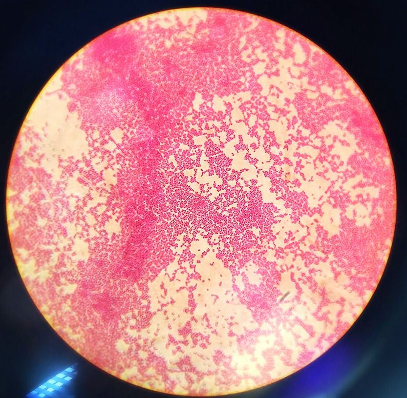
Source of image: Escherichia coli, Shahirah Shukri, UPM 1000x magnification, LM.
Differential staining, i.e gram staining:

Source of image: Gram positive Bacillus cereus, Shahirah Shukri, UPM 1000x magnification, LM.
Special staining, i.e negative staining:

Source of image: No capsule, Bacillus cereus, Shahirah Shukri, UPM 1000x magnification, LM.
Here are another examples for staining results:


LESSON 4: Microscope Observation
WHAT IS MICROSCOPE ?
For this course, we are well aware that microorganisms cannot be seen using our naked eyes. Thus, we need a helper for us to look at these amazing tiny teeny microorganisms. The helper we are talking about is microscope!
Microscope is use to enhance resolution. It is used to create a large image of the object that we cannot see with our own eyes. Generally, the greater the magnification, the greater the resolution. Magnification is the ration between the size of an image and it actual size. Resolution is the minimum distance between two objects that allows them to be seen separately. In another words, a microscope with a poor resolution, we might see only one cellular granule. But with a greater resolution microscope, it would show two granules next to each other.
If oil is put between the samples and the objective lens, the resolving power will increase. Below is the image of microscope.

Source of image : http://fileserver.net-texts.com/asset.aspx?dl=no&id=139058
MICROSCOPE COMPONENT AND ITS FUNCTION
Now, let us discuss about each part of the microscope.


Source of image: Microscope Olympus, Farhana Bajunid, UPM
HOW TO USE MICROSCOPE
Set the microscope on a flat surface. Plug’s the microscope power. First, switch on the microscope’s light course. Then, adjust the diaphragm to the largest hole diameter. In order to maximize the amount of light.
Next, make sure to rotate the nosepiece to the lowest power lens, usually 4x.
Put the slide with specimen on the stage. Then clipped onto the mechanical stage.
Adjust the coarse focus knob. Until the specimen is focused. If necessary, move the slide to the center slowly.
To have a much clearer image, adjust the small fine focus knob. Then adjust the diaphragm to control the amount of light through.
Scan the slide. To get an overview of the specimen.
Rotate the nosepiece, to the 10x objective lens for 100x magnification. Refocus the specimen and adjust the light.
LESSON 5: Movements of Microorganisms
WHAT IS CHEMOTAXIS
Microorganisms also need to move from one place to another. But the question is, how do they move? Which part that helps them to move?
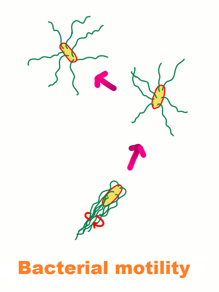
Not all microorganisms are motile. But, most motile microorganisms move by using flagella. Flagella have a hollow, rigid cylinder that is composed of a protein known as flagellin, which is attached to the basal body. In some bacteria, there is only a single flagellum, it is called monotrichous. Let's take a look at other type of microbes that has more than one flagellum.

Source of image: Izyan Fartini, UPM
A taxis is the movement of an organism in response to a stimulus such as light or the presence of food.
Chemotaxis described how the microorganisms move. It is the movement of small organisms and single cells in response to chemical signals in the surrounding environment. In chemotaxis, the microorganisms respond to the chemicals surrounding them by moving closer or further away from them. They have receptors that sensitive to the particular chemicals causing them to produce a response by using a different types of method for movement
Chemoattractants are chemicals that tend to increase the desire to approach a given chemical source. It is also known as positive chemotaxis. It is the process that occur when the microorganisms move towards the higher concentration of the chemical. While chemorepellants stimulate the microorganisms or cells to move in the opposite direction. It is called negative chemotaxis.
For example, a motile push itself from place to place by rotating its flagella. To move forward, the flagella will rotate counterclockwise and the "swims".But when flagellar rotation suddenly changes to clockwise, the "tumbles" in place and seems incapable of going anywhere. Then the begins to swim again in random direction.
The image below shown a bacterium "run" or "swim"

Source of image: Farhana Bajunid, UPM
The image below shown image of bacteria with "tumble" movement:
Source of image: Farhana Bajunid, UPM
Swimming is more frequent as the bacterium approaches a chemoattractant, which is the food. Tumbling, direction change, is more frequent as the bacterium moves away from the chemoattractant. It is a complex combination of swimming and tumbling that keeps them in areas of higher food concentrations.
LESSON 6: Treatment & Prevention of Diseases
Did you know that, microbes causing disease are not only virus and bacteria but also other types of microbes such as fungi, protozoa & helminths? That's why it is important for us to know how to prevent from getting infected and fallen sick, we need to take antimicrobial drugs which prescribed by your doctor.

Source of image: Freepik vector
Antimicrobial drugs
Antimicrobial drugs such as antibiotics are medicines that fight against bacterial infection. They either can kill the bacteria instantly or stop the growth of bacteria thus allowing the body immune system to defence & eliminate the microbes causing disease in the body. Antibiotic must be taken according to doctor’s prescription. If antibiotic is taken without the necessity, this might increase the antibiotic resistance and reduce the effectiveness of the antibiotic drugs.
Prevention
There are simple tips that can prevent infection.

Source of image: http://image.slidesharecdn.com/hand-washing-1234300301326652-1/95/hand-washing-9-728.jpg?cb=1266312727
1. Washing hands frequently & effectively
Microbes are everywhere on any inert surfaces. It is important to wash hands regularly so that the microbes on any surface that we touch will be discarded from our hands. Those microbes that temporarily resides on our hand or any surface of our body is called transient microbes. These microbes are able to enter any area that are susceptible for bacteria to enter such as nose, mouth, eyes or break of the skin and they will become opportunistic pathogen especially if the person has weak immune system. Good hand washing techniques prevent the “germs” spreading from the touch of your hands to other people or from any surfaces that you touch and then transmit to you. However, washing hand regularly without knowing to wash using proper technique is not going to remove germs on our hands effectively. The CDC recommends washing thoroughly and vigorously with soap and water for at least 20 seconds, followed by hand-drying with a paper towel. In the absence of running water, an alcohol-based hand gel or wipe can help to sanitize our hands free from germs.

Source of image: https://www.healthtap.com/topics/hand-sanitizer-kill-bacteria

Source of image: http://www.flatvision.co.uk/new-anti-bacterial-coated-monitors-for-flatvision-products-ltd/
2. Cover our mouth when sneezing or coughing

Source of image: http://www.howitworksdaily.com/wp-content/uploads/2015/11/RS32747_DSC_0193.jpg
Why covering your mouth when sneezing or coughing is important besides having personal clean hygiene? When we sneeze or coughing, water droplets secretes from our mouth and nose will release to the open air. The microbes also will spread to the air through our microscopic water droplets. When the water droplets that carry microbes reach other people, the microbes that resides on the skin will try to enter in the body. If the microbes able to breach the immune system, that people will get diseases. The current recommendation is to cover your mouth with tissue, handkerchief or if you don’t have both, cover your mouth with your arm, sleeve, or crook of the elbow, rather than not covering your mouth or using your bare hands.
3. Get vaccinated

Source of image:<a href='http://www.freepik.com/free-photo/girl-receiving-vaccine-in-the-medical-office_854541.htm'>Designed by Freepik</a>
Vaccination is one of the most reliable way to stop the infection by the microbes causing disease. Our body have different antibodies with different functions. We have T and B cells (both are adaptive immune response in third line defense) which can form into memory cells. These memory cells will recognize the antigen (microbes) and will initiate immune response if we get infected for the second time. In order to instill memory of the antigen, the body need to recognize by first antigen to the body so the memory cells will be stored. If the body encounter with the same antigen for the second time, the memory cells will initiate and defense mechanism are carry out to fight against the antigen. So, this vaccination is a process to introduce body with attenuated microbes so that the body able to store memory and thus immune system will be able to stimulate to fight against the disease causing microbes. The attenuated microbes will not cause disease or harm to the body because it is too weak to cause an action. There are never cases has been reported that these microbes can cause side effects such as autism to the body.
4. Use safe cooking and hygienic practice

Source of image: http://www.fda.gov/Food/ResourcesForYou/Consumers/ucm255180.htm
Food-borne illnesses usually arise from poor food preparation, dirty hygiene and dining habits. Microbes are always present on the food when left at room temperature.

In order to avoid spoiled from your yesterday’s food, we keep our food in the refrigerator. Refrigeration slows or stops the growth of most microbes. Promptly refrigerate foods within 2 hours of preparation. It is advisable, to use separate cutting boards for raw meats and vegetables, keep clean counter-tops, and wash all fruits and vegetables well prior to eating. Wash hands using the hand washing technique before and after food preparation.
#prokaryote #eukaryote #microscope #bacteria #coccus #bacillus #DNA #microbes #microorganisms #antibiotic #antimicrobialdrugs #pH #Temperature #Mesophiles #Psychrophiles #Thermophiles #acidophiles #neutrophiles #alkaliphiles #Oxygen #Traceelements #Anaerobic #Staining #Gramstain #Acidfaststain #Chemotaxis #Taxis #Motility #Washhands #Vaccine #Prevention


Comments